The importance of quality gynecological instruments in women’s health
Gynecology is one of the most important medical specialties in comprehensive women’s healthcare. It focuses on the prevention, diagnosis, and treatment of diseases related to the female reproductive system, as well as supporting key processes such as menstrual health, fertility, pregnancy, and menopause.
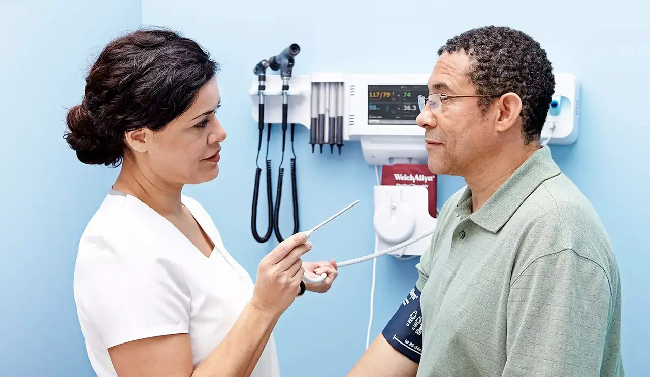
In this context, the role of the healthcare professional is fundamental, but so is the use of appropriate gynecological instruments, since precision, safety and hygiene are determining factors to guarantee optimal clinical results and a positive experience for the patient.
Technological advancements have led to the development of safer, more ergonomic, and more efficient gynecological devices, capable of improving visibility, reducing risks, and optimizing working time in consultations and operating rooms. This article offers an introduction to the world of gynecology and analyzes the importance of using high-quality equipment, as well as presenting innovative solutions available on the market.
Gynecology and its role in women’s health
Gynecology focuses not only on treating diseases but also on prevention and health education. Regular checkups, Pap smears, pelvic exams, and ultrasounds are routine procedures that allow for the early detection of pathologies, significantly improving the prognosis and quality of life for patients.
Among the most common pathologies addressed by this specialty are:
- Gynecological infections
- Endometriosis
- Uterine fibroids
- Hormonal disorders
- Polycystic ovary syndrome
- Gynecological Cancer
- Fertility Problems
For the diagnosis and treatment of these conditions, professionals need tools that provide accuracy, visibility, and safety, making the use of specialized medical instruments indispensable

Importance of high-quality gynecological instruments
The female reproductive system is anatomically complex and delicate, so any procedure requires maximum precision. High-quality gynecological instruments offer numerous benefits:
Greater safety for the patient
Using devices designed with biocompatible materials and quality standards reduces the risk of injury, infection, and complications.
Improved clinical accuracy
Ergonomic instruments with good visibility allow for more accurate examinations, facilitating early diagnosis.
Prevention of cross-contamination
Disposable instruments or those with advanced sterilization systems help minimize the risk of pathogen transmission.
Optimization of clinical time
Easy-to-use devices that do not require reprocessing reduce preparation times and improve professional efficiency.
Greater comfort for the patient
The anatomical design and ergonomics of the instruments help to reduce discomfort and anxiety during the examination.
The vaginal speculum: a key tool in gynecology
One of the most commonly used instruments in gynecological practice is the vaginal speculum, which is essential for performing pelvic examinations, cytology, sample collection, and various diagnostic and therapeutic procedures.
Benefits of the disposable vaginal speculum
In recent years, the disposable speculum has gained popularity due to advantages such as:
- Elimination of sterilization processes
- Reduction of the risk of cross-contamination
- Greater efficiency in consultation
- Improved patient experience
- Transparency of the material that improves visibility
Furthermore, the incorporation of lighting systems has revolutionized gynecological examination, providing a clearer and more precise visual field.
Technological innovation in gynecological instruments
Technology has transformed the healthcare sector, and gynecology is no exception. Devices now exist that integrate LED lighting, advanced ergonomics, and smoke evacuation systems, designed to improve safety and clinical efficiency.
In this field, specialized companies like Equimed are committed to innovative solutions that facilitate the work of healthcare professionals and improve the patient experience.
Speculum systems with LED lighting
Lighting is a determining factor in gynecological examinations. The incorporation of LED light sources in specula allows for:
- Improved visualization of the vaginal canal and cervix
- Reduction of diagnostic errors
- Greater comfort for the professional
- Reduced exploration time
- Better experience for the patient
A prime example is the KleenSpec system, which integrates bright and uniform LED lighting, providing clear viewing without the need for cables or external light sources.
These types of devices are designed to eliminate costs associated with reprocessing and contribute to infection prevention, making them an efficient option for clinics and hospitals.

Premium mirrors with smoke evacuator
In electrosurgical procedures, the generation of surgical smoke can impair visibility and pose a risk to healthcare personnel. Therefore, specula with smoke evacuators represent a significant innovation.
The Welch Allyn 590 premium series incorporates this technology, allowing:
- Smoke removal during electrosurgical procedures
- Improved visibility of the surgical field
- Risk reduction for healthcare personnel
- Prevention of cross-contamination
These solutions contribute to raising the standards of safety and efficiency in modern gynecological practice.

Current trends in gynecological equipment
The field of gynecology continues to evolve towards safer, more efficient, and patient-centered care models. Among the most relevant trends are:
Disposable Instruments
Growing concerns about safety and infection control are driving the use of single-use devices
Integrated Lighting
Integrating LED lights into instruments improves diagnostic accuracy
Advanced Ergonomics
The professional-centered design reduces fatigue and improves accuracy
Digitization and connectivity
Integration with digital systems facilitates clinical recording and patient monitoring.
Focus on the patient experience
The development of more comfortable and less invasive devices improves adherence to gynecological check-ups.

How to choose the right gynecological instruments
Selecting the right equipment is key to ensuring safe and efficient clinical practice. Some factors to consider are:
- Quality and health certifications
- Biocompatible materials
- Ergonomics and ease of use
- Compatibility with other devices
- Total cost (including reprocessing)
- Technological Innovation
- Patient Safety
Choosing specialized providers allows access to solutions designed specifically for the needs of the gynecological professional

Benefits of innovative gynecological equipment for clinics and hospitals
Investing in quality instruments not only improves clinical safety, but also provides operational and economic advantages:
- Reduction of healthcare-associated infections
- Improving staff efficiency
- Optimizing patient flow
- Greater patient satisfaction
- Competitive differentiation of the medical center
- Compliance with health regulations
These advantages make innovative gynecological equipment a strategic element for healthcare centers.

The future of gynecology: technology and safety
The future of gynecology will be defined by technological innovation and personalized care. The integration of smart devices, artificial intelligence, and advanced materials will further improve diagnostic accuracy and patient safety.
Furthermore, the focus on prevention and patient experience will continue to drive the development of more comfortable, safe, and efficient instruments, reinforcing the importance of quality equipment in gynecological practice.